Overview
CellColoc is a Python package for interactive, segmentation-based colocalization analysis in microscopy images.
The package is designed for workflows in which a user wants to:
segment a larger biological object in one channel,
segment or threshold a marker-defining structure in a second channel,
classify cells as marker-positive or marker-negative based on overlap,
optionally include a third channel for occupancy quantification or optional third-marker positivity,
inspect, refine, and export results in a reproducible way,
analyze a single microscopy channel when no colocalization step is needed and the goal is object counting, occupancy, or morphology analysis.
The package supports both 2D and 3D data, flexible segmentation backends (we support both Cellpose and classical thresholding), and a range of optional features including optional ROI-based analysis, optional global z-cropping, optional z-projection, and fast post hoc Cellpose threshold refinement.
Motivation
Many microscopy analysis scripts start as project-specific prototypes and then grow by repeated copying and adaptation. This quickly leads to duplicated core logic, inconsistent result formats, and hidden workflow drift across projects.
CellColoc addresses this by separating:
reusable core analysis functions inside the package,
project-specific configuration and execution logic inside interactive user scripts.
This keeps the analysis transparent and inspectable for researchers while still making the underlying workflow reusable across datasets and projects.
Core design
CellColoc is built around a generic channel model:
one primary
cellchannel,one primary
markerchannel,one optional third analysis channel,
an optional dedicated single-channel workflow for pure segmentation, counting, occupancy, and morphology analysis,
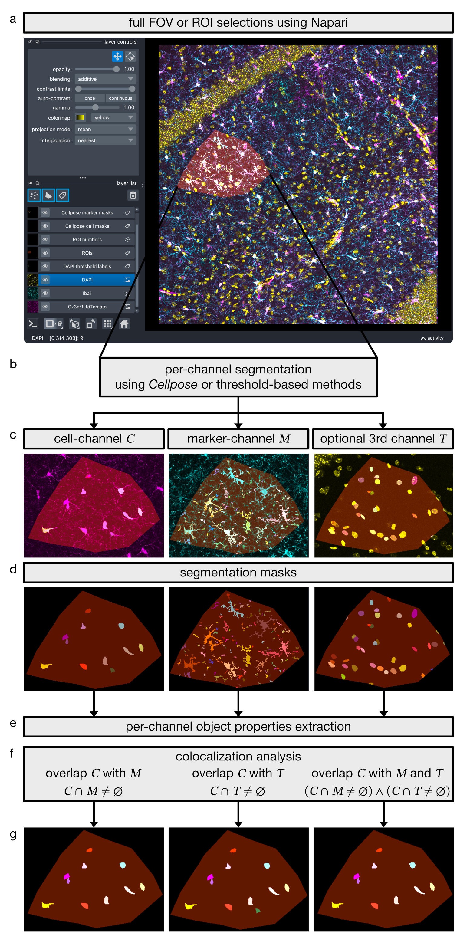
Figure 1. CellColoc workflow for object-based multi-channel colocalization analysis in microscopy images. (a) A full field of view (FOV) or user-defined regions of interest (ROIs) are selected interactively in napari. (b) Within the selected analysis region, each channel is segmented independently using either Cellpose or threshold-based methods. (c) Representative input channels for the cell channel \(C\), marker channel \(M\), and an optional third channel \(T\). (d) Corresponding per-channel segmentation masks. (e) Per-channel object properties are extracted from the segmented masks. (f) Object-based colocalization is then evaluated by testing cell-channel objects for overlap with the marker channel (\(C \cap M \neq \varnothing\)), with the optional third channel (\(C \cap T \neq \varnothing\)), or with both channels (\((C \cap M \neq \varnothing) \land (C \cap T \neq \varnothing)\)). (g) Resulting subsets of cell-channel objects classified as positive for \(M\), positive for \(T\), or positive for both \(M\) and \(T\).
Each analysis channel can use one of several segmentation backends:
cellposeotsulipercentile
This means the package is not restricted to Cellpose-only workflows. Neural network segmentation and threshold-based segmentation can be combined on a channel-by-channel basis.
Main features
CellColoc currently provides:
OMIO-based microscopy loading
automatic 2D versus 3D detection
optional voxel-size resolution from OMIO metadata
dedicated single-channel workflow for segmentation, object counting, occupancy, and morphology analysis without a colocalization step
optional ROI drawing in napari
optional whole-image analysis as a single ROI
optional reuse of saved ROI masks
ROI-wise segmentation and overlap analysis
occupancy metrics for every segmented channel
optional third-channel cell-positivity analysis
optional global z-cropping
optional global z-projection
optional prefilter chains and postfilter chains
fast Cellpose cache-based refinement
optional manual mask editing followed by table recomputation
standardized table and mask export into a
results/folder
How do we define colocalization?
CellColoc determines colocalization from segmented objects, not from raw pixel-wise intensity correlation.
For one ROI \(R\), let
\(C_i \subseteq R\) be the voxel or pixel set of cell object \(i\) in the primary
cellchannel,\(M_j \subseteq R\) be the voxel or pixel set of marker object \(j\) in the primary
markerchannel,\(M = \bigcup_j M_j\) be the union of all marker-positive pixels or voxels in that ROI.
For each segmented cell object \(C_i\), CellColoc computes the overlap with the marker segmentation as
where \(| \cdot |\) denotes the number of pixels in 2D or voxels in 3D.
In addition, the overlap fraction of the cell object is computed as
A cell is classified as marker-positive when both of the following conditions are fulfilled:
and
where \(O_{\min}\) is the minimum required overlap in pixels or voxels and
\(\tau\) is the minimum required overlap fraction. In the package, these
two thresholds correspond to the configurable parameters
min_overlap_voxels and overlap_fraction_threshold.
This means that a cell is called positive only if the overlap is both absolutely large enough and sufficiently large relative to the size of the segmented cell object.
Occupancy computation
In addition to object-based colocalization, CellColoc also computes occupancy
for every segmented analysis channel within each ROI. This includes the
primary cell channel, the primary marker channel, and, when
configured, an optional third segmented channel.
If \(S \subseteq R\) is the union of all positive pixels or voxels of one segmented channel inside ROI \(R\), then the occupancy coverage is
reported as percent coverage.
For 3D data, CellColoc can report this in volumetric form. For projected data, the same definition is applied to the analyzed 2D image.
When an optional third channel is included, it can additionally be used for:
occupancy-only analysis, for example lesion, tumor, or infiltration coverage,
separate third-channel cell positivity,
derived double-positive counts for cells that are positive for both the main marker and the third segmented channel.
Interactive workflow
The intended usage model is interactive and notebook-like:
define dataset paths and channel assignments,
choose segmentation settings,
load the analysis channels,
optionally draw or reload ROIs,
run segmentation and colocalization,
inspect results in napari,
optionally refine thresholds or masks,
export the final results.
This makes CellColoc especially suitable for exploratory but still reproducible microscopy workflows.
Where to start
We recommend to start with usage examples on this documentation website. On CellColoc’s GitHub repository, you will additionally find a folder user_scripts/ with interactive scripts that are described in the documentation and can be run cell by cell in VS Code’s interactive window or in a notebook-like environment. They are designed to be run with provided example datasets (download from Zenodo) or with your own microscopy data.
License
CellColoc is distributed under the terms of the GNU General Public License v3.0 or later (GPL-3.0-or-later).
Citation
If you use CellColoc in scientific work, please cite:
Musacchio, F. (2026). CellColoc: A Python package for interactive segmentation-based colocalization analysis in microscopy images. Zenodo. https://doi.org/10.5281/zenodo.20787509
For questions, suggestions or bug reports, please refer to the GitHub issue tracker of the CellColoc repository or contact the maintainer directly: